Corneal Foreign Body Removal: A Step-by-Step Clinical Guide for GPs
Featuring how iXope is revolutionising front-line eye care in general practice
Corneal foreign bodies (CFBs) are among the most frequent eye emergencies presenting to general practitioners and emergency departments. Timely removal is essential to avoid complications like infection, scarring, or long-term visual impairment.
This guide offers a practical, evidence-based approach to managing corneal foreign bodies, and shows how iXope - a next-generation diagnostic device - is changing the game in everyday practice, especially in remote and resource-limited settings.
Clinical Overview
-
Prevalence: CFBs account for ~30–40% of all eye-related presentations in emergency and primary care.
-
Incidence: 0.00074 – 0.00324 internationally, for Australia ~43,000 cases per year (2013 -2015)
-
Common causes: Metal (grinding), wood splinters, sand, glass, or organic matter.
-
At-risk groups: Tradespeople, rural workers, gardeners—often without eye protection.
-
Complications: Infection, corneal ulceration, rust ring (metallic FBs), and vision loss if not managed properly.
Typical Presentation
Symptoms:
-
Sudden eye pain or discomfort
-
Tearing, redness, foreign body sensation
-
Photophobia
-
Blurred vision or difficulty focusing
Clinical Signs:
-
Visible foreign body on the cornea or under eyelids
-
Fluorescein staining showing abrasion or defect
-
Rust rings (24–48 hours post-metallic FB)
-
Conjunctival injection
Step-by-Step Foreign Body Removal
1. Initial Assessment
-
Take detailed history: nature of object, timing, prior symptoms, and use of eye protection.
-
Document visual acuity first (before any intervention).
-
Apply topical anaesthetic (e.g., oxybuprocaine).
-
Use a slit lamp or equivalent magnification (like iXope) with good lighting and focal depth
-
Consider XR if ocular penetration is expected
2. Localisation
-
Use 2%fluorescein dye and blue light to identify epithelial defects.
-
Evert the eyelids to exclude FBs trapped in conjunctiva.
-
Magnification is crucial for accurate assessment.
3. Removal Techniques
-
Instil local anaesthetic (e.g. 0.4% oxybuprocaine): effective after 20 seconds , lasting up to 20 minutes
-
Consider topical pupil dilator (e.g. 1% cyclopentolate) to reduce ciliary spasm
Technique |
Indication |
|---|---|
|
Saline irrigation |
Loose surface debris |
|
Moistened cotton swab |
Lightly adherent FBs |
|
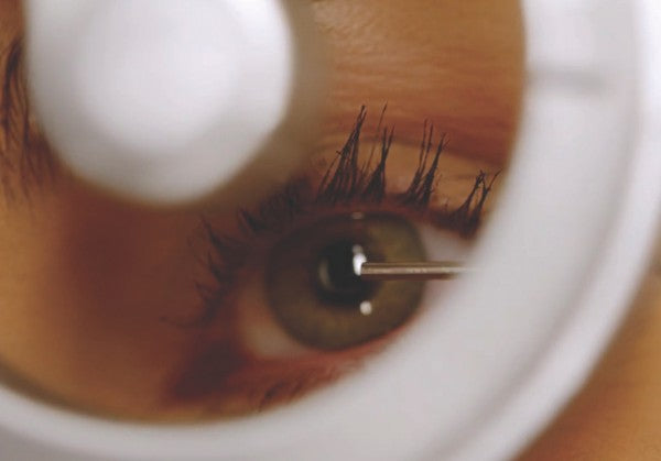
25G needle/spud |
Embedded or metallic FBs |
|
Burr |
Rust ring removal |
Always approach tangentially to the cornea. Confirm removal and recheck visual acuity post-procedure.
Follow-Up Plan
-
Antibiotic eye ointment (e.g. 0.5% chloramphenicol)
-
Consider double eye pad
-
Do not discharge with topical anaesthetics (toxicity and masking serious conditions!)
-
Mild abrasion or superficial FB: Review in 24–48 hours.
-
Rust ring or persistent symptoms: Reassess in 2–3 days.
-
Refer to ophthalmology if:
-
Vision is impaired
-
FB is deeply embedded
-
Suspected penetration of CFB
-
Incomplete removal
-
Persistent rust ring
-
Keratitis
-
Endophthalmitis
-
Persistent epithelia defects
-
Recurrent erosions
-
Paediatric or uncooperative patients
-
Infection suspected
-
Unable to tolerate in-office removal
How iXope Transforms Corneal Foreign Body Management
Traditional tools often fall short in primary care due to limited visibility, lack of documentation, or the need for referral. iXope, a portable, multifunctional diagnostic and surgical device, bridges the gap with smart features tailored for front-line care.
iXope Advantages:
-
Slit lamp–level magnification in a handheld, portable format
-
Built-in blue light for fluorescein examination of the corneal surface
-
Enables high-definition imaging and video for documentation and tracking
-
Can assist in removal of corneal foreign bodies using clear magnified view
-
Doubles as a general-purpose surgical microscope for minor procedures
-
Wireless and compact—ideal for use in remote and rural areas
-
Affordable solution for practices without access to large ophthalmic equipment
iXope in Action: sharing insights - shaping outcomes, saving time and costs
-
Helps keep patients out of hospital by enabling procedures safely in the clinic
-
Saves time for patients, doctors, and hospital triage systems
-
Reduces burden on public healthcare, saving taxpayer money
-
Captured images and video improve communication with colleagues and referrals
-
Enables streamlined follow-up, with before/after comparison of healing progress
-
Improves patient cooperation and trust through shared visual explanations
Summary: Conventional vs iXope in Corneal FB Management
Feature |
Conventional Tools |
iXope |
|---|---|---|
|
Magnification |
Basic or absent, alternatively slit lamp with good magnification |
Slit lamp–like HD optics |
|
Blue light for fluorescein |
Often Requires separate equipment |
Built-in |
|
Image capture |
Not possible or requires personal phone |
Secure, clinical-grade imaging |
|
Follow-up visual tracking |
Subjective |
Objective and image-based |
|
Use in rural areas |
Limited |
Highly portable and wireless |
|
Surgical usability |
No |
Supports minor surgical tasks |
|
Workflow efficiency |
Fragmented |
Integrated and time-saving |
Final Thoughts
iXope offers a new standard of care for general practice. With enhanced visibility, integrated imaging, and portability, it enables GPs to manage corneal foreign bodies with the confidence and precision of a specialised clinic.
Whether you’re treating patients in urban clinics or rural outreach, iXope empowers you to do more with less - and deliver better outcomes, faster.