How iXope’s dermatoscope function enhances skin cancer detection in GP practices
Skin cancer is the most common form of cancer managed by GPs and accounts for 3% of all issues managed in primary care.
Sometimes, you’re deliberately looking for it, performing a full body skin check or examining a mole that a patient is worried about. Sometimes, the patient is there for a different reason entirely but you notice a suspicious lesion and need to start investigating.
At this point, you may find yourself glancing at the clock. You can’t ignore the lesion but you know it’s going to take time to examine it, document your findings, explain things to the patient and make any necessary referrals.
iXope simplifies skin cancer checks and keeps the consultation moving. Here’s how it works.
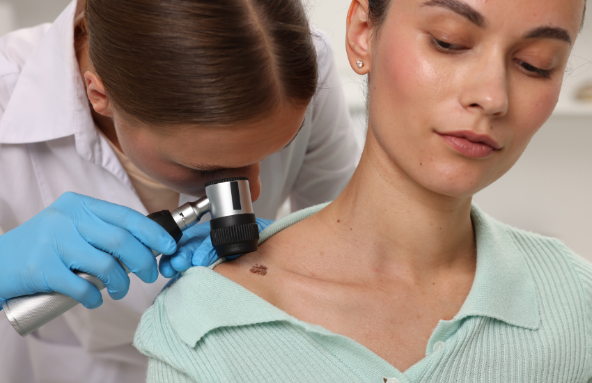
Dermatoscopy: what you can see, clearly
iXope delivers crisp, high-resolution views that make dermoscopic structures easier to interpret. Even illumination and stable focus reduce glare and artefacts, so you can evaluate pigment networks, symmetry, borders, colours and sub-surface structures rather than fighting the lighting.
Because the optics produce a consistent field of view, you can frame lesions in a repeatable way from visit to visit, improving your ability to detect subtle changes over time.
Image capture and transfer: documentation without the friction
iXope contains a built-in digital camera so you can capture images of suspicious lesions in seconds and transfer them wirelessly to your computer for the best patient care, medicolegal documentation and referrals.
No adapters, no phone juggling, no broken clinical flow.
Images land where you need them for notes, recalls and follow-up comparisons. This smooth pipeline turns longitudinal imaging into a routine part of care, so side-by-side reviews are quick, objective and easily shareable.
Referral ease: clearer letters, faster triage
Attaching a high-quality image to your referral gives dermatology colleagues an immediate visual context, aiding triage and advice.
Where teledermatology is appropriate, a clear dermoscopic image reduces back-and-forth clarification and focuses the next step – monitor, biopsy or escalate.
The same visuals also support patient conversations in the room, building their understanding of why you’re recommending a particular plan.
Time savings: in the exam and in the notes
A cable-free, point-capture-save workflow keeps appointments moving, whether you’re doing a full skin check or addressing an incidental spot at the end of an unrelated consult.
Clear images shorten written descriptions, and reliable first-time capture reduces double-handling and unnecessary re-attendance just to re-image a lesion. The result is more time for clinical reasoning and patient discussion.
Built for busy metro clinics and rural practice alike
Because iXope is compact, multipurpose, handheld and wireless, it fits crowded treatment rooms and travels easily between consulting suites, home visits or outreach clinics.
For rural and outer-metro clinics, reliable digital imaging supports local care while enabling timely escalation when malignant features are suspected.
How GPs use iXope’s dermatoscope across common skin cancer scenarios
- Dedicated skin checks: Systematic head-to-toe examination with immediate image capture for any lesion of interest.
- Incidental findings: A new asymmetric lesion spotted during an unrelated consult? Document it on the spot without extending the appointment.
- Patient-initiated “spot checks”: Quick, clear imaging when someone asks about a single mole or a changing patch.
- High-risk surveillance: Consistent, reproducible images support regular review for patients with prior melanoma, multiple dysplastic naevi, or significant sun exposure.
- Post-procedure and healing review: Visual records help confirm expected healing and identify concerning changes early.
-
Team-based care: Nurses and registrars can capture images that you review and action, improving throughput and continuity.
Small touches that make a big difference include:
- High-resolution capture to computer: Clean images ready for notes, recalls and clinical letters.
- Fast, cable-free workflow: No adapters, no downtime – just point, capture, and continue.
- Reproducible follow-ups: Same device, same framing approach, better comparisons over time.
-
Patient communication: Show and explain dermoscopic features to support informed decisions.
From first look to next step
iXope’s dermatoscope function pairs optical clarity with dependable documentation to elevate your skin cancer examination. It means you can:
- Identify lesions that warrant closer follow-up or biopsy
- Monitor suspicious lesions accurately over time
- Provide dermatologists with images that can speed advice or triage when escalation is appropriate.
Disclaimer
This information is intended for healthcare professionals.
References
-
Johns, M., O’Bryen, J., Banney, L., & Neale, R. (2024). Skin cancer prevention in Australia. Australian Journal of General Practice, 53(8), 524–527. https://doi.org/10.31128/AJGP-11-23-7010, [Accessed on 27 August 2025]