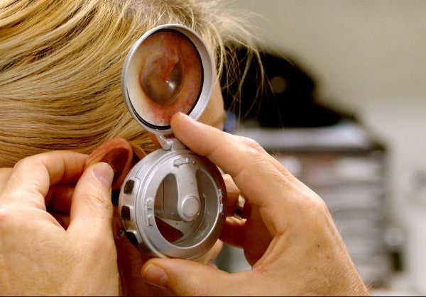
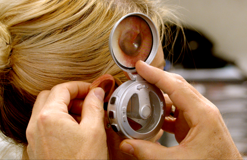
Ear Conditions Requiring Clear Visualisation of the Tympanic Membrane

What GPs Need to Know About When and How to Use MBS Item 41647
Clear visualisation of the tympanic membrane (TM) is essential for accurate diagnosis and safe management of numerous ear conditions commonly encountered in general practice. In recent years, concerns around MBS item 41647 have led many GPs to avoid using the item, despite having invested in microscopes and adopting best practices like microsuction.
Let’s break down the clinical rationale, when the item is justified, and the critical difference between complicated and uncomplicated ear wax removal — for safe, compliant, and evidence-based care.
Why Is a Clear View of the TM So Important?
A wide range of conditions require direct inspection of the eardrum. Here’s a table outlining the clinical reasons:
Condition |
Why a Clear View of the TM Is Required |
|---|---|
|
Otitis media (acute or with effusion) |
TM inspection shows bulging, retraction, redness, fluid level, or air bubbles |
|
Tympanic membrane perforation |
Size, position, and edge characteristics guide healing potential and need for ENT referral |
|
Tympanosclerosis |
Calcified plaques can mimic infection or other pathology |
|
Cholesteatoma |
Identification of attic crusting, marginal perforation, and retraction pockets requires magnification |
|
Barotrauma (e.g. post-flight, diving) |
To detect haemorrhagic TM, fluid build-up, or rupture |
|
Foreign body assessment |
Ensures TM integrity during and after removal |
|
Otitis externa with swelling/debris |
TM often hidden — need to rule out middle ear involvement |
|
Sudden hearing loss |
Differentiates between conductive and sensorineural causes |
|
Post-surgical follow-up (e.g. grommets) |
Verifies correct placement, healing, or complications |
|
Myringitis (bullous or localised inflammation) |
Requires magnified TM inspection to confirm diagnosis |
|
Eustachian tube dysfunction |
TM may show retraction, air bubbles, or fluid level |
|
TM retraction pockets |
Can progress to cholesteatoma; needs close follow-up |
|
Glue ear |
Requires visual confirmation of middle ear effusion or dull TM |
Why Use a Microscope or Endoscope?
-
Many of these conditions involve subtle or obscured TM changes.
-
Wax, swelling, pus, or canal anatomy may block otoscopic view.
-
Magnification helps detect retraction pockets, perforation edges, or early cholesteatoma.
-
Accurate assessment ensures correct diagnosis and prevents inappropriate treatment.
Clinical Tip:
If your patient presents with:
-
Otalgia
-
Discharge
-
Acute hearing loss
-
Or if you cannot visualise the TM clearly…
You are clinically justified in:
-
Performing microsuction to clear the canal
-
Conducting micro-inspection with a microscope or endoscope
-
Billing MBS item 41647, provided there is an associated ear disorder — not just simple wax
When to Claim MBS Item 41647
Below are common clinical scenarios where MBS item 41647 is justifiable:
Condition |
Why It Qualifies |
|---|---|
|
Impacted cerumen with hearing loss |
Sudden conductive loss + failed ear drops or syringing |
|
Otitis externa with canal obstruction |
Prevents TM assessment; microsuction needed |
|
Otitis media |
Requires visual confirmation of bulging TM, effusion, or perforation |
|
Foreign body |
Blocks hearing and TM view; microscope assists safe removal |
|
Cholesteatoma / canal keratosis |
Debris may block view; magnification critical |
|
TM perforation |
Need to assess margins and secondary infection |
|
SSNHL (sudden sensorineural hearing loss) |
Ruling out external or middle ear obstruction is essential |
|
Haemorrhagic otitis / barotrauma |
Blood and trauma can obscure TM — micro-inspection needed |
|
Post-op complications (grommets, mastoid) |
Sudden changes in hearing require magnified follow-up |
When NOT to Claim Item 41647
Condition |
Why 41647 Does NOT Apply |
|---|---|
|
Simple wax impaction |
No other ear disorder present — claim as standard consult |
|
Subjective hearing loss without objective findings |
No clinical justification for microscope use |
|
Using microscope just for comfort or routine |
Not billable unless a disorder is being diagnosed or treated |
The Difference Between Complicated vs Uncomplicated Wax Removal
Understanding this distinction is essential for both clinical safety and MBS compliance:
Uncomplicated Wax Removal
-
No ear disorder
-
Easily managed with irrigation or curette
-
No inflammation or TM pathology
-
Billing: Use standard consult (e.g. 23, 36, 44)
Complicated Wax Removal
-
Associated with:
-
Perforated TM
-
Otitis externa/media
-
Foreign body
-
Sudden hearing loss
-
Post-surgical canal
-
Microscope required for:
-
Visualisation
-
Safe removal
-
Assessment of TM
-
Billing: Item 41647 (if criteria met)
MBS Note TN.8.255 clearly states:
“Item 41647 applies where use of an operating microscope or endoscope is clinically necessary, such as where examination by conventional means (hand-held or spectacle-mounted auroscope) does not provide sufficient detail.
In addition, item 41647 cannot be claimed for the removal of uncomplicated wax in the absence of other disorders of the ear.
The removal of uncomplicated wax in the absence of other disorders of the ear by operating microscope or endoscope, or the removal of wax by microsuction or syringing using any visualisation method may be claimed as part of an MBS general attendance item provided all other requirements of the item have been met.”
Summary: When Can GPs Bill Item 41647?
You can bill MBS item 41647 if:
-
There is a clinical ear disorder (not just wax)
-
You use a microscope or endoscope
-
Ear drops or syringing have failed, are contraindicated, or are inappropriate
-
Make sure to clearly document the reason for performing microsuction and tympanic membrane inspection. To streamline the process, use the autofill or template feature in your EMR system.


